

2024 BIOD 152 Hematology Module 6 Exam With Answers (159 Solved Questions)
- Biology
- 18 pages
- Shared May 2025
Enhance your exam preparation with 2024 BIOD 152 Hematology Module 6 Exam With Answers, which features solved past exams to help you focus on key areas.
Preview 6 of 18 pages
See the first 6 pages exactly as they appear in the document.
Page 1 Read page 1 as text
BIOD 152- Module 7 Exam Updated 2024 What are the 2 methods of renal control mechanism? - -H+ secretion -conversion of bicarbonate When and where does H+ secretion occur? - -in response to pH of the extracellular fluid -secretes through renal filtrate Explain the conversion of bicarbonate in the renal control mechanism - -bicarbonate is replenished in the plasma by reclaiming it from the rental filtrate How is the renal control mechanism used during alkalosis? - renal collecting duct intercalated cells can secrete bicarbonate while simultaneously recovering H+ to lower the pH of the blood Normal blood serum levels - Normal pH = 7.35-7.45 Normal PCO2 = 35-45 mm (pressure of carbon dioxide) Normal HCO3- = 22-26 mEq/L (bicarbonate) When is acidosis and alkalosis disorders metabolic? - isn't caused the the pressure of CO2 in the blood When is acidosis and alkalosis disorders respiratory? - whether the cause is higher or lower CO2 pressure in the blood What can happen if there is severe acidosis? below 7 pH - central nervous system is markedly depressed causing: -coma -imminent death What can happen if there is severe alkalosis? above 7.8 pH - nervous system is markedly excited causing: -extreme nervousness -muscle contraction
Page 2 Page 3 Read page 3 as text
-convulsion -death due to the lack of breathing What is Metabolic acidosis? - -lower pH due to lower bicarbonate concentration (HCO3) cause of metabolic acidosis - buildup of acidic metabolic products such as acetic acid, lactic acid, diabetic ketosis or extreme diarrhea What is Metabolic alkalosis? - higher pH due to higher concentration of bicarbonate (HCO3) Causes of Metabolic alkalosis - Vomiting, intake of excess antacids, and constipation How does the respiratory system work to correct metabolic acidosis or alkalosis? - respiratory compensation How does the renal system attempt to correct the disorder? - renal compensation To determine if a patient has abnormal lab values (from a blood sample): - 1. acidosis or alkalosis 2. respiratory or metabolic 3. if multiple abnormal values: determine if 1 of the abnormal values is due to compensation by 1 of the systems Size and location of kidneys - -bean shaped -5 oz -between T12-L3 -right kidney lies slightly lower because of the liver What is the renal Hilus? - A depression on the medial border of the kidney What is the Renal sinus? - where the blood vessels, nerves and ureter enter or leave the kidney
Page 4 Read page 4 as text
Layers of the kidney - -Renal capsule: inner most -Adipose capsule: fatty layer -Renal fascia: outer most Inner regions of the kidney - -Renal cortex: outer portion with projections (cortical columns) -Renal Medulla: divided into sections (pyramids) -Renal Pelvis: funnel tube that connects to the ureter as it leaves the hilus -Calyces: extensions of rental pelvis that collect urine & drain into the renal pelvis -Ureter Blood Supply to Kidneys - -renal arteries come right off abdominal aorta -deliver 1200 ml of blood/ min -20% of cardiac output Explain the blood flow in the kidneys - -Renal arteries -branch into 5 segmental arteries -divide into lobar arteries -divide into interlobar arteries (pass brwn renal pyramids) -divide into arcuate arteries -divide into interlobular arteries -feed afferent arterioles -which supply glomeruli -after filtration, into efferent arterioles & drains into either peritubular or vasa recta capillaries Explain the blood flow out of the kidneys - -From capillaries, drains into interlobular veins -arcuate veins -interlobar veins -renal vein which exits kidney Nerve supply to kidney - Renal Plexus -nerve fibers follow renal arteries into each kidney -Sympathetic innervation
Page 5 Read page 5 as text
Ureters function - 2 that carry urine from kidneys into the bladder What is the Ureterovesical valves - -sphincter located where the ureters enter the base of the bladder -prevents backflow of urine Linning of ureters - -transitional epithelium: inner most -muscular: longitudinal & circular, uses contractions to propel urine -fibrous connective tissue: outer Urinary Bladder - -hollow muscular sac -receives and stores urine -urine exits via urethra Male vs female bladder - male: base of bladder in front of rectum, behind pubis symphysis. female: sits below uterus, in front of vagina. lower max cap Anatomy of the bladder - -2 urethral orifices -trigone: smooth triangular center region of the bladder Layers of the bladder - -outer adventitia: fibrous connective tissue -detrusor muscle: inner & outer longitudinal layers, middle circular layer -inner mucosal layer Why is the inner mucosal layer important to the bladder? - made up of transitional epithelium which enables expansion to absorb fluids Features of the bladder - -elastic & collapsable when empty -swells & rises in abdominal cavity -rugae -micturition What allows the bladder to extend to increase in capacity? - Rugae- inner folds
Page 6 Read page 6 as text
What is the max capacity of the bladder? How much urine causes the urge to urinate? - - 800-1000 mL -200 ml What is micturition? - the act of emptying the bladder How does micturition happen? - -detrusor muscle contracts forcing urine agains internal urethral sphincter -internal sphincter relaxes -then we control relaxing the external urethral sphincter which would result in urination What is the inability to control micturition voluntarily called? - incontinence Causes of incontinence - -infant -dementia -emotional trauma -pregnancy -Nervous system injuries What is the inability to expel urine called? - Urinary retention -catheter for treatment What causes urinary retention? - -detrusor muscle is slow to regain activity after being under anesthesia -overgrowth of prostate gland Lining of urethra - mucosal lining starts as transitional epithelium and becomes stratified columnar towards exit What is the internal urethra sphincter? - -involuntary -near bladder -keeps urethra closed
12 more pages in the full document
Unlock it once and it stays in your library, ready to chat with or turn into flashcards.
Unlock the full documentMore Biology study materials
See all Exam Guides
Test Bank for Genetics Essentials: Concepts and Connections, 3rd Edition
Biology
561 pages147downloadsFeb 2025 Past Exams
2024-2025 BIOL235 Nutrition Practice Exam with Answers (38 Solved Questions)
Biology
6 pages145downloadsFeb 2025 Exam Guides
Test Bank for Macroeconomics, 6th Edition
Biology
373 pages143downloadsFeb 2025 Exam Guides
Test Bank for Radiologic Science for Technologists: Physics, Biology, and Protection, 12th Edition (Chapters 1-40)
Biology
235 pages139downloadsFeb 2025 Past Exams
BIO1100 Anatomy and Physiology Openstax Test Bank with Answers (166 Solved Questions)
Biology
40 pages130downloadsFeb 2025 Exam Guides
Human Biology: Concepts And Current Issues, 8th Edition Test Bank
Biology
631 pages125downloadsFeb 2025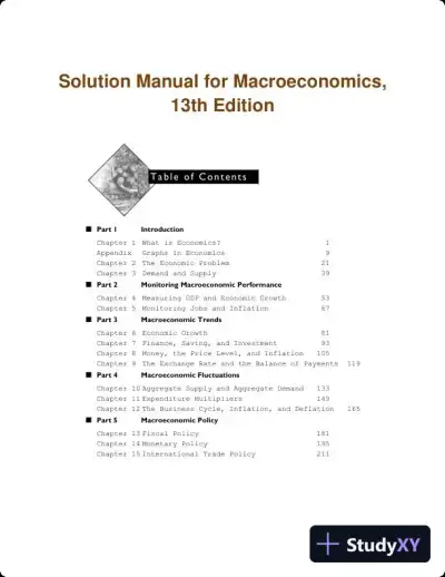 Textbook Guides
Solution Manual for Macroeconomics, 13th Edition
Biology
245 pages108downloadsFeb 2025 Textbook Guides
Solution Manual for Managerial Accounting, 6th Edition
Biology
929 pages100downloadsFeb 2025
Study this document with CramX
Once it is in your library, every tool can work from it.