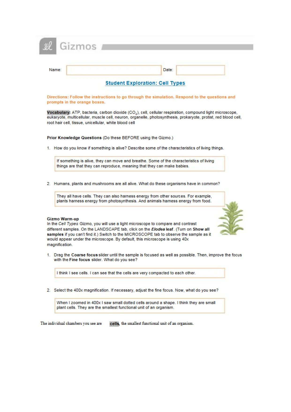
 151 GizmosName:Date:Student Exploration: Cell TypesDirections: Follow the instructions to go through the simulation. Respond to the questions andprompts in the orange boxes.Vocabulary:ATP, bacteria, cartoon dioxide (CO2), cell, cellular respiration, compound light microscope,eukaryote, multicellular, muscle cell, neuron, organelle, photosynthesis, prokaryote, protist, red blood cell,root hair cell, tissue, unicellular, white blood cellPrior Knowledge Questions {Dothese BEFORE using the Gizmo.)1.How do you know if something is alive? Describe some of the characteristicsofliving things.If something is alive, they can move and toreathe. Some of the characteristics of livingthings are that they can reproduce, meaning that they can make babies.2.Humans, plants and mushrooms are all alive. What do these organisms have in common?They all have cells. They can also harness energy from other sources. For example,plants harness energy from photosynthesis. And animals harness energyfromfood.Gizmo Warm-upIn theCeil TypesGizmo, you will use a light microscope to compare and contrastdifferent samples. On the LANDSCAPE tab click on theEtodealeaf{TurnonShow allsamplesif you can't find it.) Switc h to the MICROSCOPE tab to observe the sample as itwould appear under the microscope. By default, this microscope is using 40xmagnification.Drag the Coarse focus slider until the sample isfoeused as well as possible. Then, improve the focuswiththe Finefocusslider. What do you see?1.I thinkIsee cells.I cansee that the cells are very compacted to each other.2.Select the 40Dx magnification. If necessary, adjust the fine focus. Now, what do you see?When I zoomed in 4D0x I saw small dotted cells around a shape.Ithink they are smallplant cells. They are the smallest functional unit of an organism.The individual chambers you see arecellsthe smallest functional unit of an organism.
Preview Mode
This document has 10 pages. Sign in to access the full document!