

Computed and digital radiography notes
- Electrical Engineering & Electronics
- 6 pages
- Shared June 2025
Notes on computed and digital radiography: Digital imaging started with CT and MRI. In 1981, Fuji launched the first commercial computed radiography system. CR is a key form of digital radiography.
Preview 2 of 6 pages
See the first 2 pages exactly as they appear in the document.
Page 1 Read page 1 as text
Chapter 15- COMPUTED RADIOGRAPH? ■ *“ A cassette, similar to the cassette used in conventional radiography, encases a phosphor plate rather than film. v When x-rays strike the phosphor plate, electrons are crapped in a high energy state The latent image is stored in the grains of the phosphor in the plate. v The phosphor plate is then fed into a computer reader where it is scanned by a laser light After amplification and numbering, the light signal is digitized through a series of steps, which creates a visible image. The patient is positioned the same as in conventional radiography. COMPUTED RADIOGRAPHIC' IMAGE RECEPTOR v Photostimulable Luminescence > Some materials such as barium fluorohalide with europium (EaFEriEu or EaFLEu) emit some light promptly in the way that a scintillator does following x-ray exposure. > They also emit light some time later when exposed to a different light source. Such a process is called photostimulable luminescence (FSL). > The atoms of barium fluorobromide have atomic numbers of 56, 9, and 35, respectively, with K- shell electron bindittg energies of 37, 5, and 12 keV, respectively. > Many Compton and photoelectric x-ray interactions occur with outer-shell electrons, sending them into an excited, metastable state. Digital Imaging began ivith computed radiography (CT) and Magnetic resonance imaging (MRI). 1981, Fuji introduced digital radiography with the first commercial computed radiography imaging system Medical imaging is complemented by multiple forms of DR in addition to CR. Much of the informatian relevant to CR applies also to DR because CRis a form of DR CR& Screen-film Radiography SIMILARITIES ■ Both modalities use as the image receptor an x- ray-serisitive plate that is encased in a protective cassette. ■ The two techniques can be used interchangeably with any x-ray imaging system ■ Both produce a latent image, although in a different form that must be made visible via processing DIFFERENCE Screen-film Radiography, intensifying screen is a scintillator that emits light in response to an x- ray interaction. ■ CR, the response to x-ray interaction is seen as trapped electrons in a higher energy metastable state. CONCEPT OF COMPUTED RADIOGRAPHY The latent image in CR is held on a phosphor plate.
Page 2 Read page 2 as text
When these electrons return to the ground state., visible light is emitted. > SPSs are mechanically stable, electrostatically protected and fashioned to optimize the intensity of stimulated light Some SPSs incorporate phosphors grown as Linear filaments chat enhance the absorption of x-rays and limit the spread of stimulated emission, IMAGING PLATE The PSP screen is housed in a rugged cassette that appears similar to a screen-film cassette. * Over time, these metastable electrons return to the ground state on their own. However, this return to the ground state can be accelerated or stimulated by exposing the phosphor to intense infrared light from a laser—hence the term photostimulable luminescence from a photostimulable phosphor [PSP]. * The PSP. barium fluorohalide, is fashioned similarly to a radiographic intensifying screen. LIGHT STIMULATION-EMISSION Sequence of events engaged in producing a PSL: 1. EXPOSE When an x-ray beam exposes a Photostimulable Phosphor [PSP], the energy transfer results in excitation of electrons into a metastable state. Approximately 50% of these electrons return to their ground state immediately, resulting in prompt emission of fight 2 STJMEZLA77OM The finely focused beam of infrared light with a beam diameter of 50 to IDO pm is directed at the PSP. As laser beam intensity increases, so does the intensity of the emitted signal Note that as die laser beam penetrates, it spreads. The amount of spread increases with PSP thickness. The diameter of the spatial resolution of die ( STORAGE PHOSPHOR SCREENS [STS] Screens for latent image formation through metastable electrons. Appears white because the small PSP partides scatters liglit excessively. - called turbid > PSP particles are randomly positioned throughout a binder. e laser beam determines imaging system
4 more pages in the full document
Unlock it once and it stays in your library, ready to chat with or turn into flashcards.
Unlock the full documentMore Electrical Engineering & Electronics study materials
See all Textbook Guides
Solution Manual for Microwave Circuit Design A Practical Approach Using ADS, 1st Edition
Electrical Engineering & Electronics
121 pages154downloadsFeb 2025 Exam Guides
Power System Analysis And Design, 6th Edition Test Bank
Electrical Engineering & Electronics
40 pages132downloadsFeb 2025 Textbook Guides
Solution Manual For Electronics Fundamentals: A Systems Approach, 1st Edition
Electrical Engineering & Electronics
278 pages93downloadsFeb 2025 Textbook Guides
Electronic Commerce, 12th Edition Solution Manual
Electrical Engineering & Electronics
134 pagesMay 2025 Textbook Guides
Solution Manual for Signals and Systems using MATLAB , 3rd Edition
Electrical Engineering & Electronics
538 pagesMay 2025 Document
HW4_Solution
Electrical Engineering & Electronics
17 pagesFeb 2026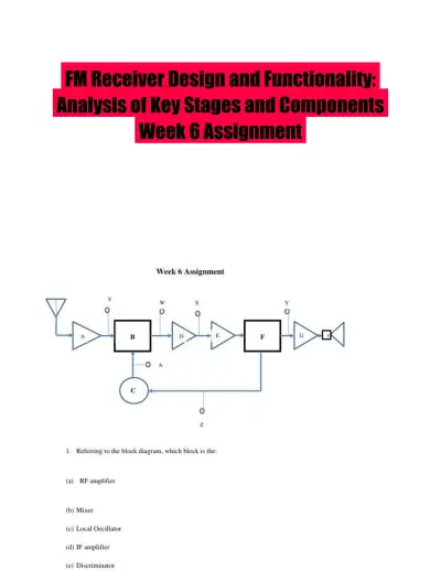 Solved Assignments
FM Receiver Design and Functionality: Analysis of Key Stages and Components Week 6 Assignment
Electrical Engineering & Electronics
5 pagesMay 2025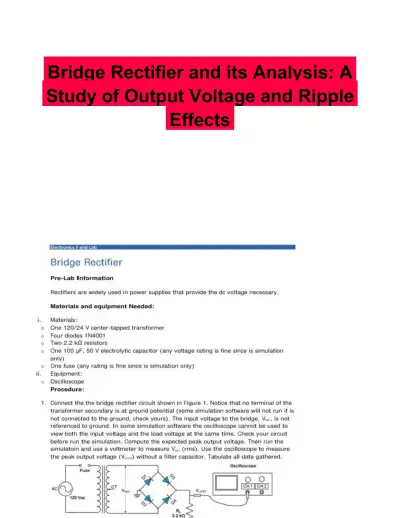 Solved Assignments
Bridge Rectifier and its Analysis: A Study of Output Voltage and Ripple Effects
Electrical Engineering & Electronics
3 pagesMay 2025
Study this document with CramX
Once it is in your library, every tool can work from it.